

AI-Powered Detection of Hypochromic Red Blood Cells in Peripheral Blood Smears
DOI:
https://doi.org/10.14740/aicm32Keywords:
Deep learning image processing, Computer-assisted algorithms automation, Laboratory anemia, Iron-deficiency erythrocytes, Abnormal blood chemical analysis, Pathologists, Validation studiesAbstract
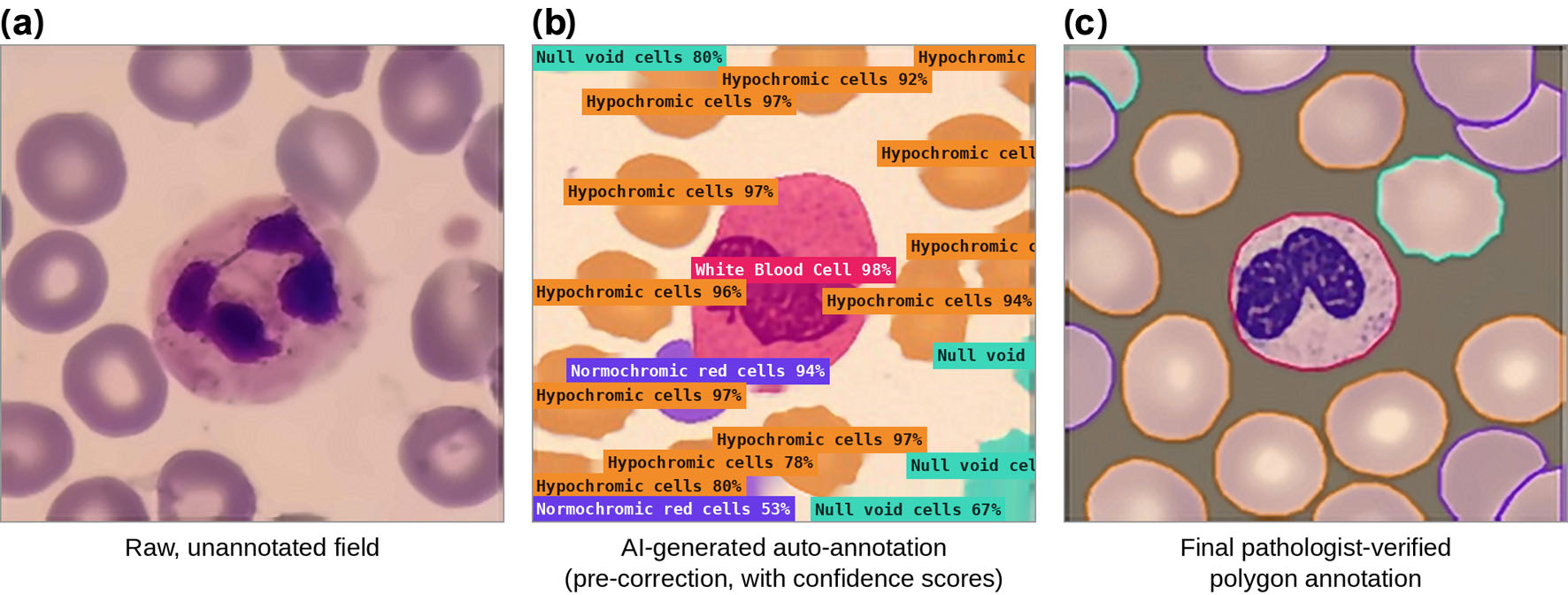
Background: Manual peripheral blood smear (PBS) review for hypochromic red blood cells (RBCs) in iron deficiency anemia (IDA) screening is laborious and subjective. This retrospective, secondary data-analysis development and validation study evaluated a computer vision deep neural network to automate hypochromic RBC detection from PBS images using a pathologist-in-the-loop semi-automated annotation workflow.
Methods: From April to June 2026, a pathologist manually annotated a 100-image subset of the public TXL-PBC dataset (870 images in total). A preliminary model generated label-assisted annotation for the remaining 770 images, which were verified by the pathologist and trained using the Receptive Field Detection Transformer (RF-DETR) instance segmentation network. Samples with a hypochromic-to-total RBC ratio > 20% were flagged as morphologically suggestive of IDA (a morphological screening criterion, not a stand-alone etiological diagnosis). External validation was performed using 600 confirmed IDA cases from the ANERBC 2 dataset.
Results: The RF-DETR network achieved mAP@0.5 = 90.0%, precision = 86.0%, recall = 85.9%, and F1 = 89.5%. The validation workflow correctly identified 594/600 confirmed IDA cases (99% sensitivity), establishing hypochromic RBCs as a dominant, statistically separable, and artefact-resistant marker. The semi-automated annotation pipeline reduced dataset curation time by 62.1%.
Conclusion: The pathologist-assisted RF-DETR network enables accurate, scalable IDA screening from PBS images with significantly reduced annotation burdens. Strong performance on an independent external cohort supports its utility as a morphological screening adjunct for IDA, to be interpreted alongside clinical and laboratory iron indices rather than as a stand-alone diagnostic test.
Published
Issue
Section
License
Copyright (c) 2026 The authors

This work is licensed under a Creative Commons Attribution 4.0 International License.