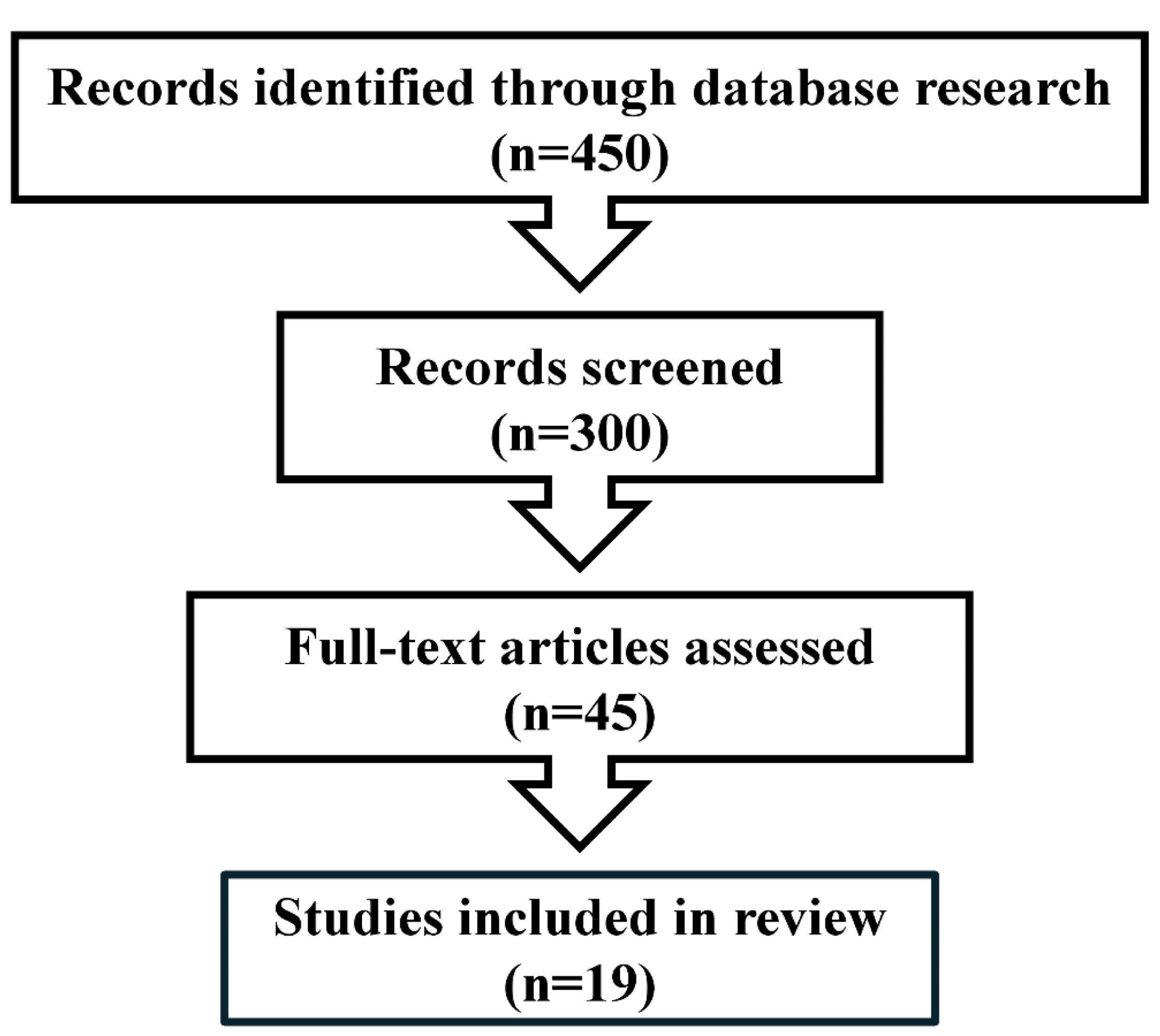
↓ Figure 1. PRISMA flow diagram.
| AI in Clinical Medicine, ISSN 2819-7437 online, Open Access |
| Article copyright, the authors; Journal compilation copyright, AI Clin Med and Elmer Press Inc |
| Journal website https://aicm.elmerpub.com |
Review
Volume 2, March 2026, e16
Artificial Intelligence in Colposcopy: Diagnostic Performance, Clinical Applications, and Future Directions
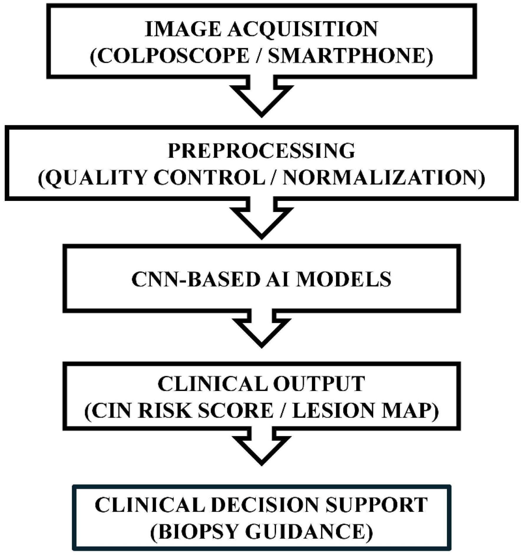
Figures
Table
| Study | Year | Study design | Dataset | AI method | Target outcome | Performance |
|---|---|---|---|---|---|---|
| AUC: area under the receiver operating characteristic curve; CNN: convolutional neural network. | ||||||
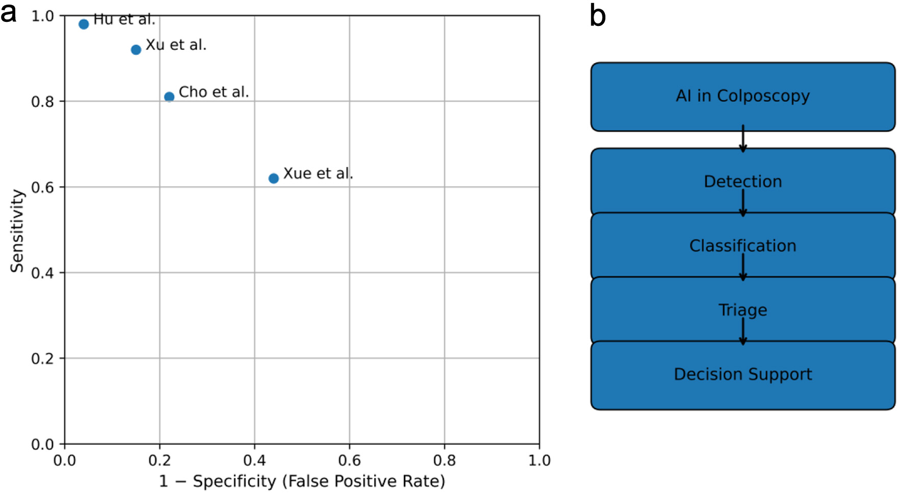
| Hu et al [5] | 2019 | Retrospective | 9,406 images | CNN | CIN2+ vs. ≤ CIN1 | Sensitivity 94.0%, specificity 88.0%, AUC 0.96 |
| Song et al [6] | 2020 | Retrospective | 7,531 images | Deep CNN | CIN2+ detection | Sensitivity 91.3%, specificity 80.0%, AUC 0.93 |
| Xue et al [7] | 2020 | Retrospective | 6,763 images | CNN + attention | CIN2+ detection | Sensitivity 98.0%, specificity 62.0% |
| Zhao et al [8] | 2022 | Retrospective | 3,673 images | CNN | High-grade CIN | Sensitivity 86.2%, specificity 78.6% |
| Liu et al [1] | 2024 | Meta-analysis | 33 studies | Multiple models | CIN2+ detection | Pooled sensitivity 93.0%, specificity 85.0% |